


High above the reach of the swamp’s Panicum grasses, a tiny petrified ant clenches tightly to the end of a thin stem. Though only a few millimeters in length and completely shrouded by water droplet, a single stalk erupting from the side gives away its identity. The culprit was an Ophiocordyceps fungus, a fascinating insect pathogen that manipulates its host’s behavior to survive and propagate. A fungal spore first comes into contact with an ant, and if left untouched, it will expand into infective hyphae that penetrates the exoskeleton slowly but surely, secreting enzymes to aid its descent. Once the host’s external surface is breached, the fungus proliferates within the body, securing a foothold for its next daunting move. During this time period, the ant behaves somewhat normally, going about its foraging duties to fulfill its role in the colony. But one day, things start to go south. The ant begins exhibiting erratic behaviors, rendering it incapable of feeding or carrying food reserves, and the convulsions knock it from its arboreal path to the forest floor. Here, the ant is isolated from the colony, preventing other uninfected ants from taking matters into their own hands to destroy the premature fungus.
By this time, the fungus has fully occupied the ant’s body cavity, having grown into the interstitial spaces surrounding muscle fibers and nervous tissue. Ophiocordyceps will then secrete metabolites and enterotoxins that manipulate its host’s behavior into climbing upwards and biting down viciously into a stem or leaf vein. During the ascent, solar cues are thought to play a role in the infected ant’s positioning and orientation, optimizing the light and humidity conditions for the developing fungus. Remarkably, once the biting behavior is elicited, the fungus will atrophy the ant’s mandibular muscles as hyphae secure the mummified husk, affixing the host in place. The legs will also flex around the substrate, anchored by the spiderweb-like hyphae, making it difficult to become dislodged. Over the course of several days, a single stalk (stroma) will emerge from the back of the head along with two fruiting bodies (ascoma) that become increasingly inflated. Finally, spores will be released into the air, falling to land and infect another unfortunate insect passerby. Near ant colonies, Ophiocordyceps can sometimes be observed in high densities, and the ants will avoid traversing those locations to prevent infection. So far, I’ve refrained from anthropomorphisms in this post, but it’s fun to think of ants avoiding these so-called graveyards due to “fear of the dead.”
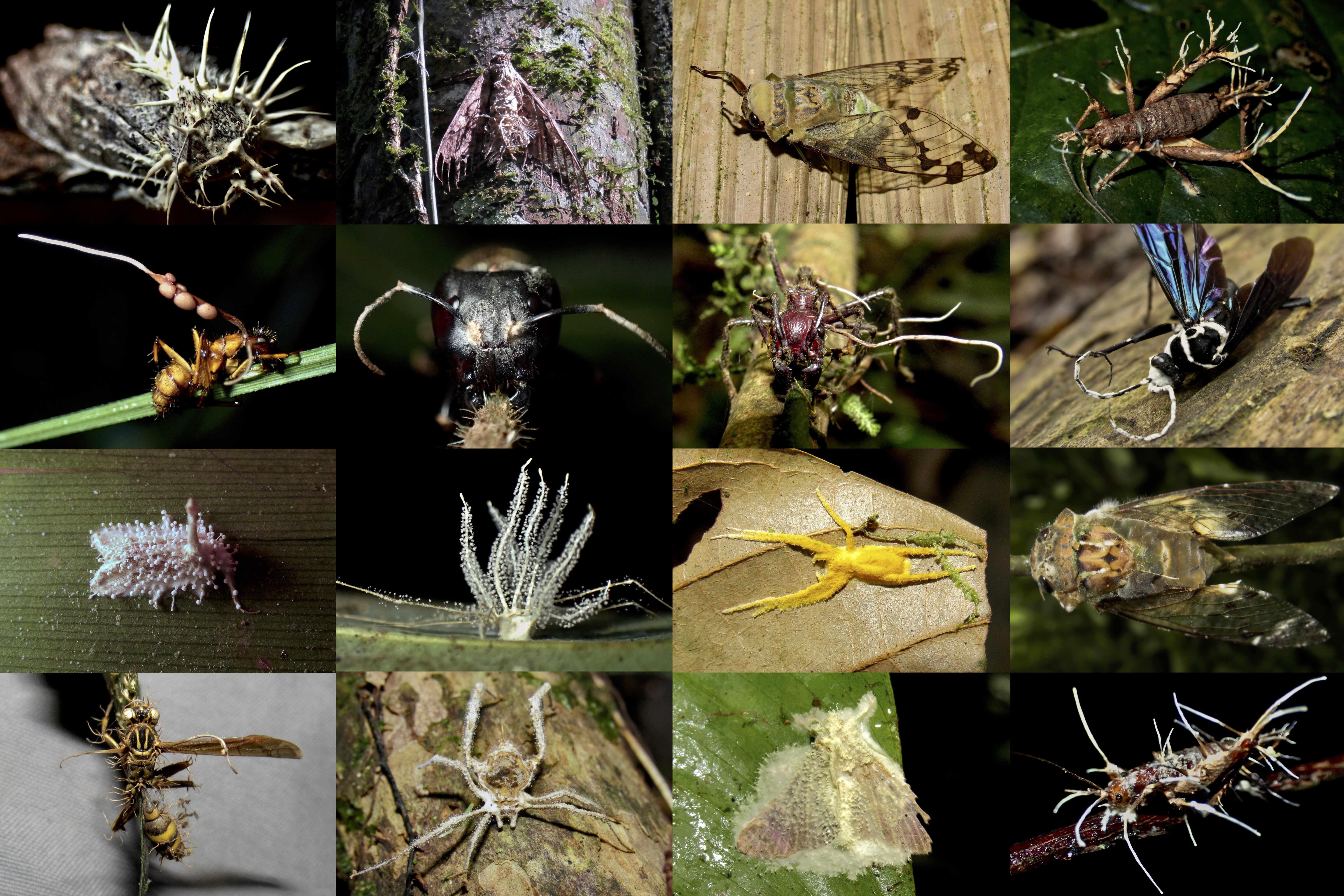
The fungus pictured above was found on a compact carpenter ant (Camponotus planatus). Although its appearance represents a classical example of Ophiocordyceps, there is high variation in fungal morphology across the genus, which likely comprises a complex of hundreds of species, each co-evolved with a particular host species. Below are more than a dozen examples of entomopathogenic fungi I’ve observed over the years, illustrating a snapshot of their incredible diversity.


Although not a fungus, another pathogenic organism called the horsehair worm (Nematomorpha) takes a similar manipulative approach to achieve its life cycle. Extremely tiny nematomorph larvae are first ingested by insects as they drink from standing bodies of water. After entering the insect’s digestive tract, larvae will feed within the gut, leaving vital organs and tissues intact. Development may take up to several months, and once mature, the nematomorph will alter its host’s behavior to wander restlessly until they find water. Infected insects do not seek water specifically, but once water is detected, hosts will fling themselves onto the water’s surface. The adult will then emerge, usually from the abdomen, and lay millions of microscope eggs in gelatinous strings. In at least some species, there may be intermediate hosts, in which a semiaquatic insect first acquires the parasite, then transfers it to a predatory terrestrial arthropod when preyed upon. Nematomorphs tend to be monochromatic pale brown or black, so the orange color here is unusual. Given that both host and parasite were frozen in place, it’s likely the caterpillar died prematurely, and the nematomorph desiccated while attempting to exit.
