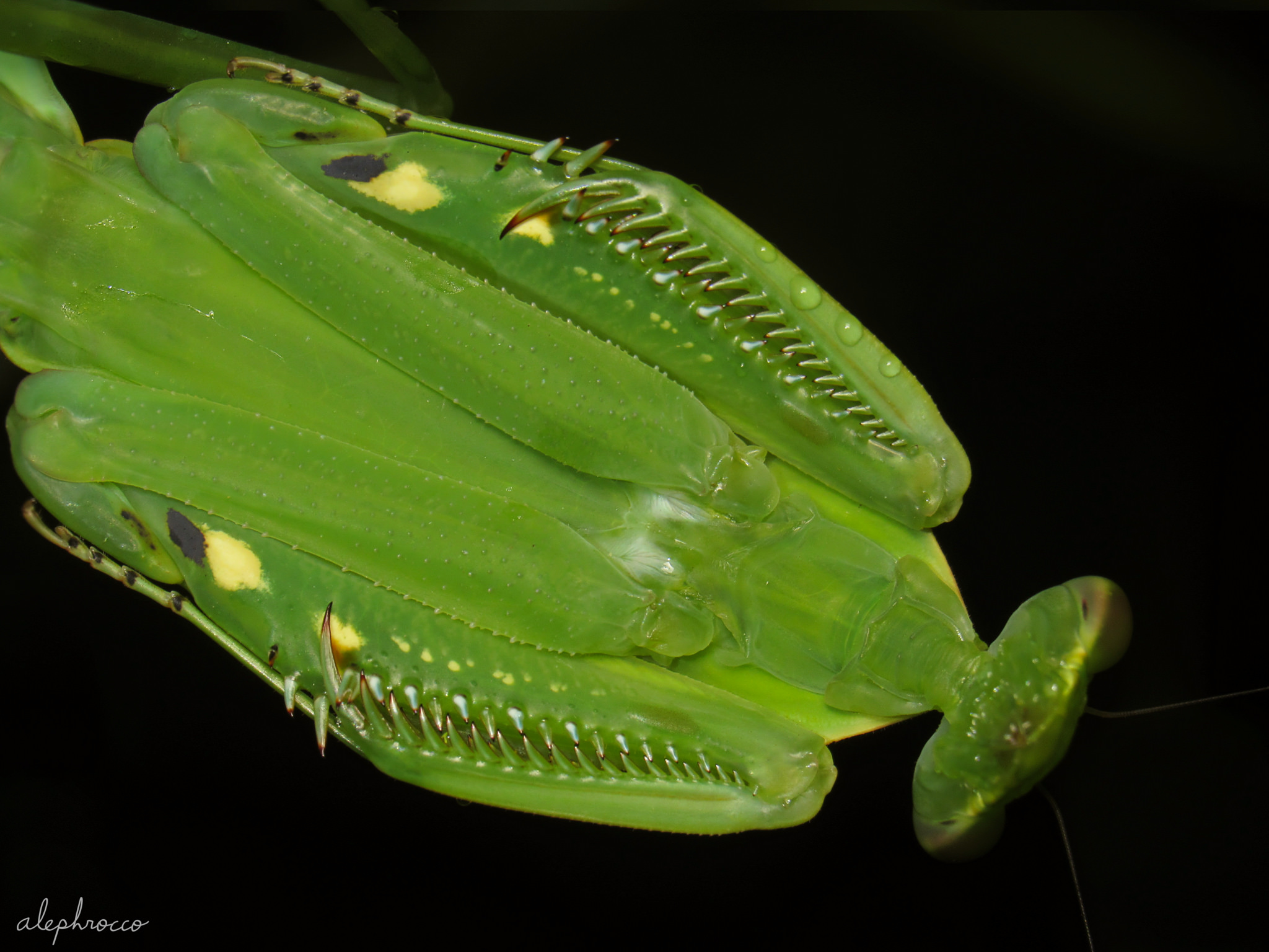
Striking symmetry in a subadult Macromantis sp., apposing her raptorials to the pronotum and flattening her body defensively. In this dorsal view you can see intricate internal structures through her semitransparent cuticle, including the slender alimentary canal and branching tracheae. To ingest food mantids chew up prey and relay the particles from the pharynx into the oesophagus. Pulsating muscular contractions (peristalsis) move the food down this thin channel into the crop prior to enzymatic digestion and excretion. Observing mantids hunt and feed is a fascinating process; it’s bizarre to watch prey gradually transform into a narrow string of nourishment, pumped down the canal in a rhythmic manner.
Inside the raptorial forearms lies a single long tube connected to tiny capillary-like structures. These are actually tracheoles, part of an insect’s tracheal system for oxygen delivery to the body’s tissues and organs. Tracheae are invaginations of cuticular cells, so the innermost layer is thickened in a circular or spiral arrangement (called taenidia), which prevent the structure from collapsing. In addition to the cuticle, tracheae are also shed during the molting process. Next time you find a cicada or cricket molt take a peek inside to see the inverted network of tubes! Tracheae terminate at the body’s surface in valvular openings called stigmata or spiracles. Breathing occurs by taking in air through the thoracic spiracles and expelling air through the abdominal spiracles (see my footage of another Costa Rican mantid, Antemna rapax: https://youtu.be/lTyhyijvURA). Gas exchange and oxygen delivery is likely a limiting factor for insect body size, so hyperoxic atmospheric environments in the Paleozoic may have enabled arthropod gigantism. Nevertheless, extant mantids of this robust genus and others such as Phasmomantis can attain over ~13 cm in length.
Photographed in situ [1]